Dr. Michael Garcia
Dr. Michael Garcia

PhD, 1999 Mayo Clinic College of Medicine
Molecular and cellular control of nerve development and disease
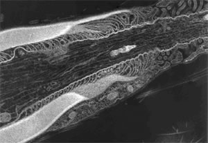
Transmission electron micrograph of an axon isolated from the 5th lumbar ventral root of an adult mouse. This image shows the transition from compact myelin (on left) to node of Ranvier. The paranodal loops indicate the end of the compact myelin and the beginning of the node of Ranvier. Within the axon, both neurofilaments and microtubules are visible.
Specification of axonal diameter is a key component of motor system development and functioning, as axonal diameter is major axonal property that specifies the velocity of electrical signal conduction in the nervous system.
The diameter of myelinated axonal segments (internodes) is increased relative to the diameter of unmyelinated axonal segments (Nodes of Ranvier). Compare the diameter of the left side of the axon in the EM to the diameter on the right side of the image. Formation of compact myelin and axonal accumulation of neurofilaments are both required for axonal diameter specification. However, it is unclear how axons and myelinating cells interact to determine axonal diameter. One proposed mechanism is that myelinating cells signal the axon resulting in alterations to the neurofilament network that support radial growth of the axon.
We are, also interested in the pathogenesis of neurodegenerative diseases such as Charcot-Marie-Tooth (CMT), amyotrophic lateral sclerosis (Lou Gehrig's Disease) and spinal muscular atrophy.
We generate genetically modified mice to investigate the role of the neurofilament network in establishing axonal diameter and to determine the mechanisms of disease pathogenesis in CMT2E.