George Smith
PhD, 1970 Harvard University
Molecular imaging of cancer through phage display
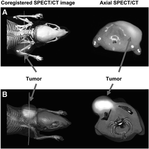
The University of Missouri is a world leader in the growing field of “molecular imaging,” which seeks to develop a new generation of agents for early detection and localization of cancers in the body. Molecular imaging differs from MRI, CAT scans, and other current imaging technologies in that the cancer cells are imaged specifically. This is accomplished by attaching a radioisotope to a molecule that binds specifically to the cancer cells and not to other tissues in the body. As an example, the picture shows a mouse into which human melanoma cells were grafted; the melanoma cells have formed a tumor at the mouse’s shoulder. A radioactive imaging agent specific for melanoma cancer cells was injected into the mouse, and a few hours later the mouse was imaged in two ways: by single photon emission computed tomography (SPECT) to detect the radioactivity (bright white area), and by a conventional CAT scan to delineate mostly the skeleton. The two images are superimposed; the superimposed image on the right is a computed section through the mouse at the line in the superimposed image on the left. You can see how the radioactive tumor-binding molecule allows the tumor to be seen much more easily than in the non-tumor-specific CAT scan. My lab’s role in the University’s molecular imaging program is to use the phage display technology developed by our group to discover novel tumor-binding molecules for common cancers, and to create easier, more powerful ways of using tumor-binding molecules for imaging.
Member, National Academy of Sciences 2020
Nobel Prize in Chemistry 2018
Promega Biotechnology Research Award, America Society for Microbiology 2007
Elected Fellow, AAAS 2001
University of Missouri Curator’s Professor 2000